
Showing posts with label Anatomy. Show all posts
Showing posts with label Anatomy. Show all posts
Thursday, June 7, 2012

Thursday, April 26, 2012
Brain - Overview
Procencephelon = Telencephelon (cerebrum) + Diancephelon (epithalamus, thalamus, hypothalamus)
Mesencephelon - Midbrain
Rhombencephelon - cerebellum , pons, medulla oblongata
Brain stem - below cerebrum, infront of cerebellum - midbrain, pons, medulla oblngata
Limbic system - primitive centre deep inside temporal lobe.
Collection of nerve cell bodies in PNS - ganglia
Collection of nerve cell bodies in CNS - Nuclei
Mesencephelon - Midbrain
Rhombencephelon - cerebellum , pons, medulla oblongata
Brain stem - below cerebrum, infront of cerebellum - midbrain, pons, medulla oblngata
Limbic system - primitive centre deep inside temporal lobe.
Collection of nerve cell bodies in PNS - ganglia
Collection of nerve cell bodies in CNS - Nuclei
Saturday, April 21, 2012
Tongue

Development -
Copula - hypobranchial eminence


Papilla

Innervation

The tongue receives its blood supply primarily from the lingual artery, a branch of the external carotid artery and lingual veins which drain into internal jugular vein.
Thoracic cage
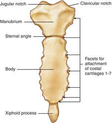
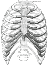
- Humans have 24 ribs (12 pairs). The second - seventh sets of ribs, known as "true ribs", are directly attached to the sternum through the costal cartilage.
- the first three (eighth, ninth, and tenth rib) have their cartilages attached to the cartilage of the rib above
- the last two (eleventh rib and twelfth rib) are free at their anterior extremities and are termed floating ribs or vertebral ribs because they connect neither with the sternum nor with another rib.
Kidney
Relations of Kidney -
the right kidney is related to the liver, duodenum, ascending colon or right colic flexure, and small intestine.
The left is related to the spleen, stomach, pancreas, descending colon or left colic flexure, and small intestine. Posteriorly, the kidneys are related to rib 12 and the diaphragm, psoas major, quadratus lumborum, and transversus abdominis
the right kidney is related to the liver, duodenum, ascending colon or right colic flexure, and small intestine.
The left is related to the spleen, stomach, pancreas, descending colon or left colic flexure, and small intestine. Posteriorly, the kidneys are related to rib 12 and the diaphragm, psoas major, quadratus lumborum, and transversus abdominis
Salivary gland histology
- The submandibular gland is a compound tubuloalveolar gland.
- It is surrounded by a capsule of moderately dense connective tissue, from which septa divide the gland into lobes and lobules.
- The secretory endpieces consist of a more or less spherical mass of cells called an acinus (pl. acini).
- The cells of the acini appear triangular in sections, with their apex directed toward the lumen, and their base resting on a basement membrane.
- They secrete their product in a merocrine fashion into the lumen.
- Contractile cells called myoepithelial cells or basket cells lie between the basement membrane and the plasma membrane of the secretory cells.
- Myoepithelial cells posess many actin-containing microfilaments, which squeeze on the secretory cells and move their products toward the excretory ducts. They can sometimes be identified in standard histological preparations.
- Acini can be either serous or mucous.
- The secretion of serous cells is thin, watery and proteinaceous.
- Serous cells have a rounded nucleus and secretory granules in their cytoplasm.
- They are joined near their apical surfaces by junctional complexes.
- Mucous cells secrete a viscous, glycoprotein-rich product, which is stored as mucinogen granules.
- The nuclei are typically flattened against the base of the cells (unless the cells have just discharged their contents, in which case they look more like serous cells).
- Mucous cells typically look pale and empty in standard histological sections, because their granules are lost during preparation.
- Its mucous acini are quite frequently capped with a serous demilune, a crescent of serous cells around one or more of their surfaces.

Types of Ducts
- There are three types of ducts in the submandibular gland: intercalated, secretory (aka striated) and excretory ducts.
- Intercalated ducts are slender ducts continuous with the terminal acini, and lined with flat, spindle-shaped cells. They secrete bicarbonate ion into and absorb chloride ion from the acinar product.
- Secretory ducts have eosinophilic cuboidal to columnar cells with basal striations. These result from infoldings of the basal membranes in which are found many mitochondria. Secretory ducts resorb sodium and secrete potassium
- Both intercalated and secretory ducts are found within the parenchyma of the gland and are therefore intralobular ducts.
- The largest ducts are the excretory ducts. They are found in the connective tissue septa, and are therefore interlobular ducts.
- They ultimately connect with the oral cavity.
- Their epithelium is variable, it can be simple cuboidal, stratified cuboidal, stratified columnar or pseudostratified.
- Near the oral cavity, it becomes stratified squamous.
- Excretory ducts do not change the secretory product.
Credit:http://www.courseweb.uottawa.ca/medicine-histology/english/gastrointestinal/Salivary.htm
Cells in Brain
Ependymal cells - line brain ventricles
Schwanna cells - around axon of nerve cell in peripheral nervous system and is derived from neural crest cells
Schwanna cells - around axon of nerve cell in peripheral nervous system and is derived from neural crest cells
Blood Supply of Brain

Anterior communicating arteries - most common aneurysms- run on top
Middle - run on sides laterally - most common stroke
Posterior - run underneath or ventrally

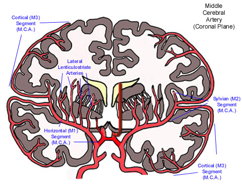
Middle cerebral artery runs on lateral surface of cerebrum. - most common in stroke esp lenticulostriate nucleus
Note: Middle meningeal - most common sub dural hematoma and a branch of maxillary artery. one of three branches supplying meninges - anterior, middle and posterior,
Anatomy of a muscle cell
Epimysium - outisde entire muscle
perimysium- around bunch of muscle fibres
Endomysium - connective tissue sheath around each muscle fibre
Sarcolemma - plasma membrane of each muscle fibre.
each muscle fibre is made of a myofibrils.


Muscle Triad - T - tubule which is an invagination of sarcolemma along wih 2 terminal cisternae ( they are just enlarged areas of sarcoplasmic reticulum storing calcium)

Sliding filament theory
Wednesday, April 18, 2012


Tuesday, April 17, 2012
Umbilical blood supply

umbilical vein - placenta to liver with oxy. blood - ligament teres of liver in adults
ductus venosus - venous shunt connects liver to Inf vena cava - ligamentum venosum in adults
formane ovale - opening btw left and rt. atria - bypasses pulm circulation - closes at birth and becomes fossa ovalis.
ductus arteriosis - between pulm trunk and aorta - bypasses pulm sirc - ligamentum arteriosum in adults
umbilical arteries - de0xy blood from fetus to placenta - lateral umbilical ligaments in adults
Trachea
pseudostratified ciliated columnar epithelium cells
commences at the lower border of the larynx, level with the sixth cervical vertebra, and bifurcates into the primary bronchi at the vertebral level of thoracic vertebrae T4/5. at the sternal angle or angel of Louis
commences at the lower border of the larynx, level with the sixth cervical vertebra, and bifurcates into the primary bronchi at the vertebral level of thoracic vertebrae T4/5. at the sternal angle or angel of Louis

Para nasal Sinus openings
above the superior concha and posterior to it - sphenoidal sinus opening
some posterior ethmoidal cells - superior meatus
maxillary sinus (hiatus semilunaris) , frontal sinus, anterior and middle ethmoidal air cells (ethmoidal bulla) - middle meatus
naso lacrimal duct opening - inferior meatus

some posterior ethmoidal cells - superior meatus
maxillary sinus (hiatus semilunaris) , frontal sinus, anterior and middle ethmoidal air cells (ethmoidal bulla) - middle meatus
naso lacrimal duct opening - inferior meatus

Ganglia of cranial nerves
Facial nerve - geniculate ganglion
Smilunar / gasserian / trigemina ganglion - sensory nucleus of trigeminal nerve
Smilunar / gasserian / trigemina ganglion - sensory nucleus of trigeminal nerve
parasympathetic ganglia of head and neck
CN 3 - Ciliary ganglia - Preganglionic axons from theEdinger-Westphal nucleus travel along the oculomotor nerve and form synapses with these cells. Thepostganglionic axons run in the short ciliary nerves and innervate two eye muscles:
- the sphincter pupillae constricts the pupil, a movement known as Miosis. The opposite, Mydriasis, is the dilation of the pupil.
- the ciliaris muscle contracts, releasing tension on the Zonular Fibers, making the lens more convex, also known as accommodation.
CN 7 - Sub Mandibular ganglion -
- Preganglionic parasympathetic fibers from the superior salivatory nucleus of the medulla oblongata, via the chorda tympani and lingual nerve, which synapse at the origin of:
- Postganglionic parasympathetic fibers to the oral mucosa and the submandibular and sublingual salivary glands.They are secretomotor to these glands.
CN - 7 Pterygo Palatine Ganglion -
Pre ganglionic - nervus intermedius (a part of the facial nerve) through the greater petrosal nerve.
Post ganglionic - trigeminal nerve to the mucous membrane of the nose, soft palate, tonsils, uvula, roof of the mouth, upper lip and gums, and upper part of the pharynx. It also sends postganglionic parasympathetic fibers to the lacrimal nerve (a branch of the Ophthalmic nerve, also part of the trigeminal nerve) via the zygomatic nerve, a branch of the maxillary nerve (from the trigeminal nerve), which then arrives at the lacrimal gland.
CN 9 - Otic ganglion - lesser petrolar nerve from inferior salivatory nucleus to parotid gland from glosso pharyngeal nerve throught auriculo temporal.
Subscribe to:
Posts (Atom)